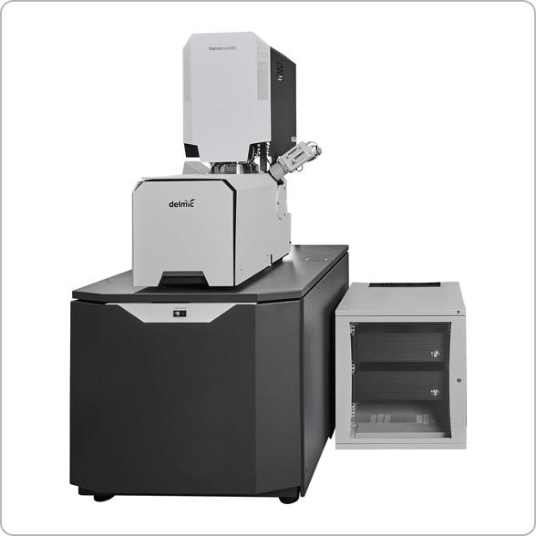
[custom_headline type=”left” level=”h1″ looks_like=”h2″]FAST-EM[/custom_headline]
Ultra-fast automated multibeam electron microscope
FAST-EM is an ultra-fast automated multibeam electron microscope (EM) designed to make complex and large EM projects simple and efficient. Thanks to its automated acquisition, this high-throughput system is ideal for imaging large or multiple samples for quantitative analysis.
Delivering powerful insights while keeping the workflows simple, this system allows users to shift their focus from microscope operation to data analysis.

FAST-EM system receives 2022 Microscopy Today Innovation Award
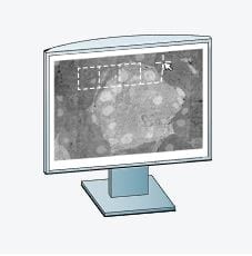
FAST-EM can be used to explore cell architecture, the interaction of neuronal circuits, and the analysis of any biological material. It is extremely beneficial for large volume 3D imaging, large scale 2D imaging and, in general, as a tool that can significantly speed up daily microscopy work.
Key benefits
Image faster
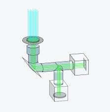
High acquisition speed by using 64 electron beams and short dwell times.
Focus on data analysis
Leave the system to automatically acquire complex datasets without constant supervision.
Achieve high sustained throughput
Minimize the overhead during imaging with robust automation.
Get the details and the big picture
Collect nanoscale detail while retaining larger context of the sample.
Workflow at glance
The reliability of the microscope and the software allow the operator to leave the system running without constant babysitting.
Overcoming challenges in large-scale imaging projects with FAST-EM
Webinar Fast Imaging: High throughput imaging with the FAST EM system
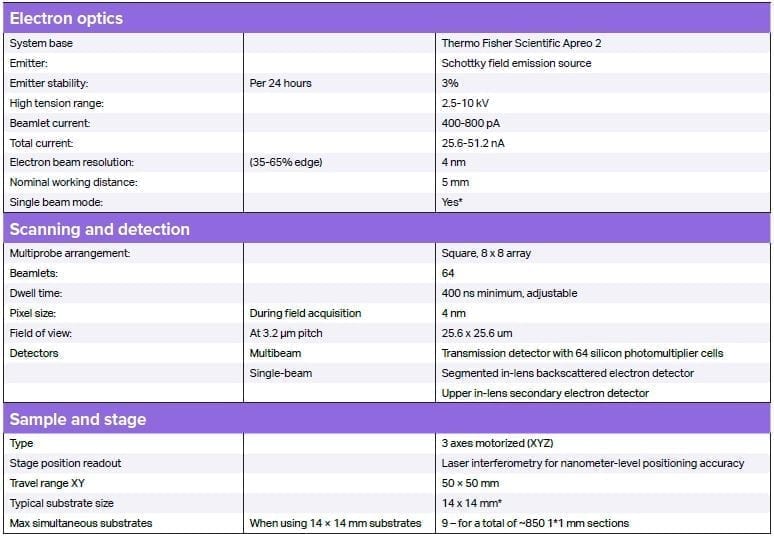
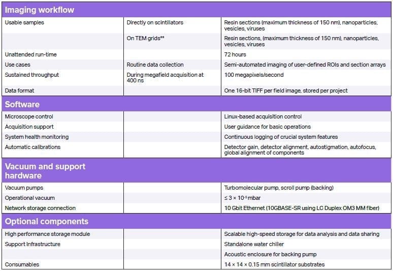
Spécifications du système